|
Design
Details
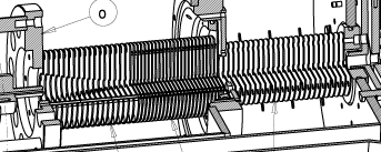
The electrospray ion mobility mass spectrometer
is set up on a steel frame cart that holds the vacuum chambers and two
diffusion pumps. A cross sectional view of the stainless steel vacuum
chambers and their interior is shown in the figure below.
The 12 in. O.D. main chamber (a) contains the ion mobility
cell (f) and is pumped by a 10 in. diffusion pump (Edwards, Sussex,
England, 2000 L/s). The 6 in. O.D. detector chamber (b) is pumped by a
6 in. diffusion pump (Edwards, Sussex, England, 700 L/s). The electrospray
source is isolated from the main chamber by two stages of differential
pumping using two mechanical pumps (E2M40, Edwards, Sussex, England) attached
to the two elbows (c) on the source flange (d).
Ions are sprayed from an external needle (not
shown) and enter the vacuum system through a capillary that feeds the
ion funnel (e). The ions are funneled and guided
through two stages of differential pumping and subsequently injected into
the drift cell (f). Ions that exit the cell are mass selected in the quadrupole
mass analyzer (g) and finally counted with a conversion dynode/CEM
detection system (h & i).
A more thorough description of the different components of the apparatus
is given in ref. [1] and in the following sections:

Top: Cross
sectional view of entire instrument as viewed from the top. Bottom: Perspective
cross sectional view of source, funnel, and cell. (a) &
(b) vacuum chambers, (c) pump ports, (d) source flange, (e) ion
funnel, (f) drift cell, (g) quadrupole mass analyzer, (h) conversion dynode,
(i) detector, (j) capillary heating block, (k) insulator, (l) funnel first
section, (m) funnel second section, (n) funnel third section, (o) funnel
flange, (p) hat flange, (q) second pump stage, (r) cell body, (s) cell
end cap, (t) ceramic ring, (u) guard rings, (v), (w),
& (x) ion optics.
|
Electrospray
The
electrospray source consists of two major components: the needle
containing the electrospray solution and the capillary that acts
as the vacuum interface. Both metalized glass needles (PicoTip,
New Objective, Cambridge, MA, 1.2 mm O.D., 2 mm tip) and hypodermic
stainless steel needles (0.004 in. O.D., ~70 mm I.D.) glued into
stainless steel tubes (1/16 in. O.D. x 0.010 in. I.D.) are used
as spray tips. The needle is mounted on an x,y,z translation stage.
The glass needles serve as nanospray tips (the needle is the liquid
reservoir) and are typically operated without any backpressure.
The hypodermic needles are fed by a syringe pump with typical flow
rates of 20-50 mL/h. Nanospray appears to be ideal for quick experiments
and for peptide work while the syringe pump setup appears to be
the method of choice for proteins. The high voltage on the spray
needle is typically +500-2000 V with respect to the capillary for
nanospray and ~2 kV for positive ion electrospray. The capillary
(0.010 in. I.D.) is 3.0 in. long and is inserted into an aluminum
block (j) which can be heated to 250 °C. The aluminum block
is mounted to a heat resistant PEEK insulator (k), which is in turn
mounted to the source flange (d).
top
|
|
Ion
Funnel
 The
ion funnel is the interface between the ESI source and the ion drift
cell located in the high vacuum chamber. It is a high transmission
RF ion guide device that has two functions. First, it compresses
the divergent stream of ions leaving the capillary down to a small
diameter. Second, it can move the ions from the source region to
the drift cell without the use of high acceleration fields, thus
avoiding high energy ion-neutral collisions. The
ion funnel is the interface between the ESI source and the ion drift
cell located in the high vacuum chamber. It is a high transmission
RF ion guide device that has two functions. First, it compresses
the divergent stream of ions leaving the capillary down to a small
diameter. Second, it can move the ions from the source region to
the drift cell without the use of high acceleration fields, thus
avoiding high energy ion-neutral collisions.
Our ion funnel was modeled after the original
designs of Smith and coworkers [2]. In our design
there are three individually tunable sections (l, m & n in the
figure on top). The first two sections are
stacks of 18 and 24 lenses (0.030 in. thick, 2.00 in. O.D.), respectively.
The lens holes in the first stack decrease parabolically from 0.87
to 0.14 in. diameter; those in the second stack decrease down to
0.10 in. diameter. The third section consists of 25 lenses (0.030
in. thick, 1.60 in. O.D., 0.16 in. I.D.). The lenses in the first
two sections are stacked on six ceramic tubes and mounted on the
funnel entrance side to a plastic flange (o). Flange (o) is mounted
to the source flange (d) allowing easy removal for cleaning.
The first 18 lenses in the funnel (with
the largest orifices) are spaced 0.100 in. apart using ceramic spacers.
These lens hole diameters are nearly identical with those reported
in ref. [2]. As the lens orifice diameter decreases
to a value comparable to the lens spacing, however, our trajectory
calculations carried out using SIMION [3] indicated
that ion transmission drops sharply. In other words, the funnel
works best when the space between the lenses is smaller than the
lens orifices. Also, the orifice size must be larger than the lens
thickness to avoid a field free region inside the lens. These effects
were found independently in Smith's lab as well.[4]
With this in mind, we decided to stack the lenses in section two
(m) closer together and to avoid small orifice diameters altogether.
Both of these goals were achieved by using 1/16 in. thick "O"
rings to space lenses 18 through 42. Since the orifices of lenses
19 through 26 decrease linearly from 0.14 in. to 0.10 in. while
lenses 27 through 42 all have 0.100 in. diameter orifices, the orifice
diameters are always greater than the lens spacing of 0.06 in. The
"O" ring spacers not only provide a closer lens spacing
but also prevent radial pumping of gas out of the lens stack. This
second effect has two consequences. First, conductance of the viscous
gas flow into the next pumping stage (q in the figure
on top) is greatly reduced because the flow is through a quasi
tube rather than a single orifice. Second, the ions are embedded
in a directed flow of gas into the next pumping stage, which should
increase ion transmission.
The final section of the funnel (n) is mounted
to the hat flange (p) and is located in the next pumping stage (q).
The first 16 lenses are again spaced by 0.100 in. ceramic spacers,
while the last 9 are spaced by 3/32 in. "O" rings to decrease
the pumping conductance into the following chamber.
The effect of pressure on the funnel operation
is somewhat uncertain. Previous reports [2,4]
and our SIMION modeling both indicate that collisions are important
in constraining the ions to the center of the funnel and reducing
their kinetic energy. In the present instrument, the funnel operates
in two pressure zones: the pressure in sections 1
& 2 (directly after the capillary)
is ~0.2 Torr; section 3 is ~0.02 Torr. Efficient ion trapping clearly
occurs in both sections, thus we can say that pressures between
0.02 and 0.2 Torr are adequate for funnel operation. Increasing
the pressure in the first sections (above 0.2 Torr) with N2
did not improve the funnel performance.
Three 1 MHz RF generators with individually
adjustable amplitudes are used for the three sections of the ion
funnel. For each section two outputs are provided, 180° phase
shifted from each other. In addition, three individually controllable
DC drift voltages are provided as well, the voltage of each section
floating on top of the previous section's potential. The appropriate
drift voltage is applied to the first and last lens of each section,
with the individual lenses being fed by a 1 MW resistor chain. The
RF is applied to the lenses via 1000 pF capacitors, alternating
the (+) and (-) outputs. The last two lenses in the funnel are independently
tunable focusing lenses and do not carry any RF.
The DC applied to section one (2.23 in.
long) is typically 10-50 V, the peak-to-peak RF voltage 100-200
V. The corresponding values for section two (2.26 in. long) are
30-50 V DC and 100-200 V RF and for section three (3.11 in. long)
1-5 V DC and 50-150 V RF. The voltage between the last lens of section
three and the drift cell entrance orifice determines the ion injection
energy. It is adjustable from 0-200 V and is typically 20-60 V.
Higher injection energies can be used to effect structural changes
and/or fragmentation of the ions, if desired.
The next to the last lens in the funnel
can be pulsed to gate the continuous ion beam from the ESI source.
A voltage high enough to stop the beam is applied to close the gate.
Since ions cannot readily escape radially due to the RF trapping
nor can they exit back into the source due to the DC ramp applied,
they accumulate near the exit of the ion funnel. The gate is opened
by pulsing the lens potential down to the normal voltage for approximately
10 μs. For cell drift times in the range
of 100 μs to 1 ms a pulsing repetition
rate of 1-10 kHz is typically used. The number of ions per second
counted after the quadrupole mass filter is approximately the same
in continuous and pulsed ion beam mode, indicating a near 100% trapping
efficiency in the ion funnel.
top
|
|
Drift
Cell
The
drift cell used in this experiment is very similar to those described
previously.[5] It consists of a near cubic copper
body (3.5 x 3.5
x 2.0 in., "b" in the figure
below), that can be heated by electrical heaters and cooled
with a flow of liquid-nitrogen cooled nitrogen gas, a copper end
cap (d) with separate temperature control, and a ceramic ring (h)
that separates the end cap from the body. The drift field in the
cylindrical interior of the cell body is provided by the entrance
(f) and exit plates (i) (0.005 in. thick, 0.5 mm orifice) held in
place by guard rings at the same potential (0.057 in. thick, 0.600
in. I.D.) and by four intermediate guard rings (e) (0.115 in. thick,
0.600 in. I.D.) equally spaced and mounted on six ceramic rods (j).
A precision 1 MW resistor chain connects the rings. The total drift
length from entrance to exit plate is 4.503 ± 0.002 cm. The
cell temperature is variable from 80 K to above 800 K.
Typical operating pressures are 4-5 Torr
with drift voltages ranging from 2-20 V/cm yielding conditions within
the low field limit.[6] Higher pressures and
voltages (up to 1000 V across the cell) are possible, however this
requires smaller cell orifice holes.
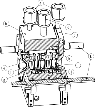
FIGURE:
Perspective cross sectional view of the temperature controlled drift
cell after Kemper and Bowers' design.[1] (a)
cooling line, (b) cell body, (c) buffer gas inlet, (d) cell end
cap, (e) drift guard ring, (f) ion entrance hole, (g) ion focusing
lens, (h) ceramic ring, (i) ion exit hole, (j) ceramic rod holding
guard rings, (k) ceramic rods holding cell assembly.
|
|
Mass
Analyzer and Detector
Ions
exiting the drift cell enter a 0-4000 amu quadrupole mass filter
("g" in the figure on top, ABB Extrel,
Pittsburgh, PA) followed by an off-axis conversion dynode (h). Particles
leaving the conversion dynode are detected by a channel electron
multiplier ("i", K &
M Electronics, West Springfield MA). The TTL signal pulses
from the preamp are collected with a multi-channel scaler board
(MCSplus, EG&G Ortec, Oak Ridge, TN). The MCS is equipped with
a voltage ramp generator, which is used to scan the quadrupole mass
analyzer.
top
|
|
Electronics
The
electronic units necessary to control the ESI source, the ion funnel,
the drift cell, and the ion optics (v) in front of the drift cell
(three lenses plus x/y-steering), (w) in front of the quadrupole
(three lenses plus x/y-steering), and (x) in front of the detector
(two lenses plus x/y-steering) were built in-house (the labels v,
w, and x refer to the figure on top). The
quadrupole mass filter employed here cannot be floated significantly
and was thus fixed at ground potential. The voltage on the drift
cell exit orifice then determines the ion energy along the quadrupole
axis; the cell entrance voltage floats on top of the exit orifice
potential and determines the drift potential; the end of the ion
funnel floats on the cell entrance potential and determines the
ion injection energy. Similarly, the three sections of the ion funnel
float on top of each other and the ESI capillary on top of the funnel.
The electrospray needle is the last element and is referenced to
the capillary. This stacking arrangement is necessary since any
change in voltage in the series (in the cell drift potential, e.g.)
must be tracked by all the preceding potentials to maintain a constant
ion formation and injection environment. If the individual lens
supplies were individually referenced to ground, any change in one
lens would require resetting all previous lens voltages.
The potentially high voltages in the ion
funnel with respect to ground and the relatively high pressure in
the ion funnel make the system prone to discharging. To avoid discharges
no grounded metal parts are present inside the ion funnel vacuum
chamber and the vacuum flange, support rods, and the support flange
for the ion funnel were made of plastic.
top
|
References
- Wyttenbach,
T.; Kemper, P. R.; Bowers, M. T. Int. J. Mass Spectrom. 2001,
212, 13-23.
- Shaffer,
S. A.; Prior, D. C.; Anderson, G. A.; Udseth, H. R.; Smith, R. D. Anal.
Chem. 1998, 70, 4111-4119.
- Dahl,
D. A. Int. J. Mass Spec. 2000, 200, 3-25. SIMION,
Version 6.0, Scientific Instrument Services, Inc., Ringoes, NJ.
- Tolmachev,
A. V.; Kim, T.; Udseth, H. R.; Smith, R. D.; Bailey, T. H.; Futrell,
J. H. Int. J. Mass Spectrom. 2000, 203, 31-47.
- a) Kemper,
P. R.; Weis, P.; Bowers, M. T. Int. J. Mass Spectrom. Ion Proc.
1997, 160, 17-37. b) Kemper,
P. R.; Bowers, M. T. J. Am. Soc. Mass Spectrom. 1990,
1, 197-207.
- Mason, E. A.; McDaniel, E. W. Transport Properties of Ions in Gases;
Wiley: New York, 1988.
top
|